خطوات إجراء الخزعة
نعني بالجراحة الدماغية المصوبة، هو الوصول الى أي بقعه أو كتله أو مكان بالدماغ بعد تحديد الاحداثيات الثلاثة لذلك الهدف عن طريق ارسال ابره، أو قطب كهربائي، أو انبوبه تصريف، أو إدخال منظار أو أشعه مركزه الى الدماغ دون احداث اذى للأنسجة الدماغية المحاذية.
وتتم هذه الجراحة بإجراء الخطوات التالية
- تركيب جهاز الجراحة الستيريوتاكتيك (Frame Stereotactic) الثلاثي الأبعاد على رأس المريض باستخدام التخدير الموضعي
- اجراء تصوير للدماغ بالمرنان المغناطيسي الثلاثي الأبعاد مع الجهاز المثبت على الرأس واحتساب الإحداثيات الثلاثة بواسطة الكومبيوتر
- نقل المريض الى غرفة العمليات أو مركز الأشعة المصوبة بالجاما نايف لأجراء ما يلزم للحالات التأليه:
- الخزعة الدماغية (Stereotactic Biopsy)
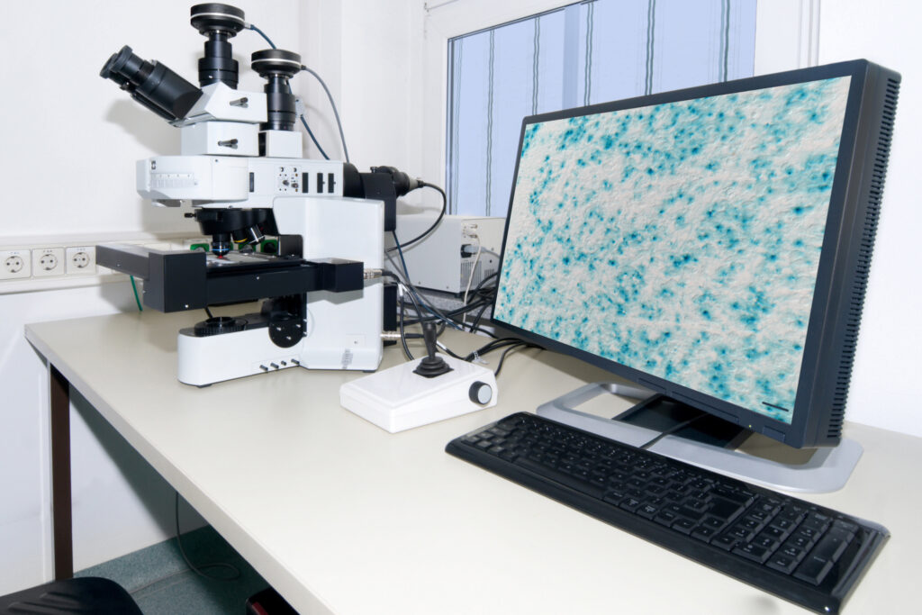
وفي هذه الحالة ندخل ابره الخزعة عبر فتحه صغيره بالجمجمة باستخدام التخدير الموضعي ونوجهها الى الورم الدماغي حسب الإحداثيات الثلاثة المحسوبة، ونقوم بأخذ الخزعة من الورم وارسال العينة الى المختبر لدراستها نسيجيًا ومخبرياً لمعرفة نوع الورم ودرجته، دون اللجوء الى فتح الدماغ. - تصريف دمل داخل عمق الدماغ (Stereotactic Brain Abscess Drainage)
في حالة وجود دمل بعمق الدماغ يصعب استئصاله جراحيًا. نقوم بزراعة انبوبة تصريف الدمل الدماغي لتصريف الصديد، وإجراء الزراعة اللازمة لمحتويات الدمل بالمختبر وحقن الدمل بالمضادات الحيوية. - تصريف أكياس سوائل دماغيه (Stereotactic Brain Cyst Drainage)
- الخزعة الدماغية (Stereotactic Biopsy)
- زراعة انبوبه دائمة داخل الورم القحفي البلعومي ومعالجته
(Stereotactic Catheter and Ommaya Reservoir Implantation For Cystic Craniopharyngioma)
يعتبر الورم القحفي البلعومي (Craniopharyngioma) من أخطر الأورام التي تظهر بالأطفال وبعض الكبار. وعادة الاستئصال للورم بالجراحة المجهرية غير مكتمل نظراً لموقعه الحساس والتصاقه بالدماغ والأماكن الحساسة، مما قد تتسبب هذه الجراحة التقليدية بالمضاعفات الخطيرة والاضطراب الهرموني وعودة نمو الورم. أما بالجراحة المصوبة الثلاثية الأبعاد، فإننا ندخل انبوبة تصريف دائمة للمحتويات السائلة للورم ونوصل الأنبوب بخزان صغير مزروع تحت فروة الرأس.
ونقوم بعد ذلك بسحب تجمع السائل من داخل الورم وعلى فترات متتالية مما يؤدي الى تخفيف الضغط الناتج على الأعصاب البصرية وقاع الدماغ. وبعد ذلك نحقن الورم بعلاج انترفيرون (Interferon) أو ماده سائله مشعه أو علاج كيماوي وذلك لقتل الورم بمهده أو استكمال معالجته بجهاز الجاما نايف.
أو ماده سائله مشعه أو علاج كيماوي وذلك لقتل الورم بمهده أو استكمال معالجته بجهاز الجاما نايف. وقد أثبت هذه الطريقة لعلاج الورم القحفي البلعومي نقاعتها وتفوقها على الجراحة المجهرية التقليدية ودون تعريض المريض لعمليات فتح الرأس ومضاعفاتها.
الجراحة المصوبة الوظيفية لعلاج مرض الباركنسون والحركات اللاإرادية Stereotactic Functional Neurosurgery For Parkinson`s Disease and Movement Disorders
الجراحة الشعاعية المصوبة بالجاما نايف لعلاج أمراض- وأورام الدماغ بدون جراحه Gamma knife Radiosurgery For Brain Diseases and Tumors

